
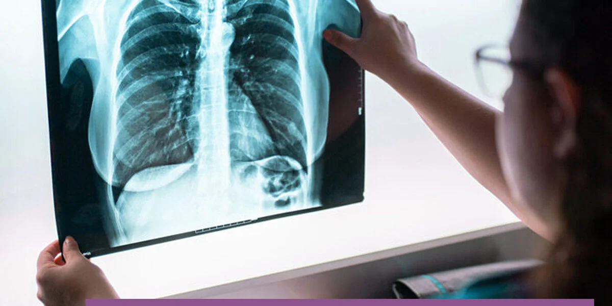
Oncology Imaging
- 4 Sections
- 7 Lessons
- 1 Quiz
- 16h 20m Duration
Front End Development
Designing Section
Bonus
Quiz
Oncology Imaging is a comprehensive course designed to provide healthcare professionals with a strong foundation in the principles, applications, interpretation, and clinical relevance of imaging in cancer care.
The course examines the role of diagnostic imaging throughout the cancer journey, including screening, early detection, diagnosis, staging, treatment planning, response assessment, surveillance, recurrence detection, and evaluation of complications. Learners will develop an understanding of how different imaging modalities are selected according to the suspected cancer, clinical question, anatomical region, and stage of disease.
Core modalities covered include plain radiography, ultrasound, mammography, computed tomography (CT), magnetic resonance imaging (MRI), positron emission tomography (PET), PET/CT, PET/MRI, nuclear medicine imaging, bone scintigraphy, and image-guided procedures. The course also introduces important concepts such as contrast-enhanced imaging, radiologic terminology, standardized reporting, RECIST response assessment, radiation safety, and imaging limitations.
Emphasis is placed on the ability of healthcare professionals to understand and clinically correlate imaging findings rather than independently replace specialist radiologic interpretation. Learners will explore how imaging findings integrate with clinical assessment, histopathology, biomarkers, molecular testing, and multidisciplinary tumor-board decision-making.
The course is particularly valuable for oncology nurses, physicians, radiographers, pharmacists, laboratory professionals, cancer researchers, and other healthcare professionals seeking advanced foundational knowledge in cancer imaging.
Want to submit a review? Login
Related Courses
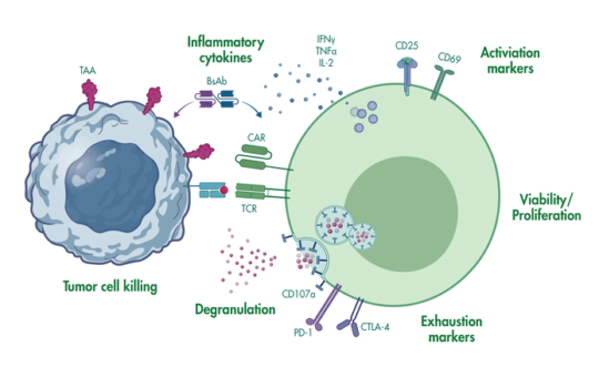

Precision Oncology & Cancer Genetics
Foundations of Precision Oncology
Cancer Genetics & Genomics
Molecular Biomarkers
Oncology Laboratory Practicals
Oncology Laboratory Fundamentals
Haematology Assessment
Clinical Chemistry